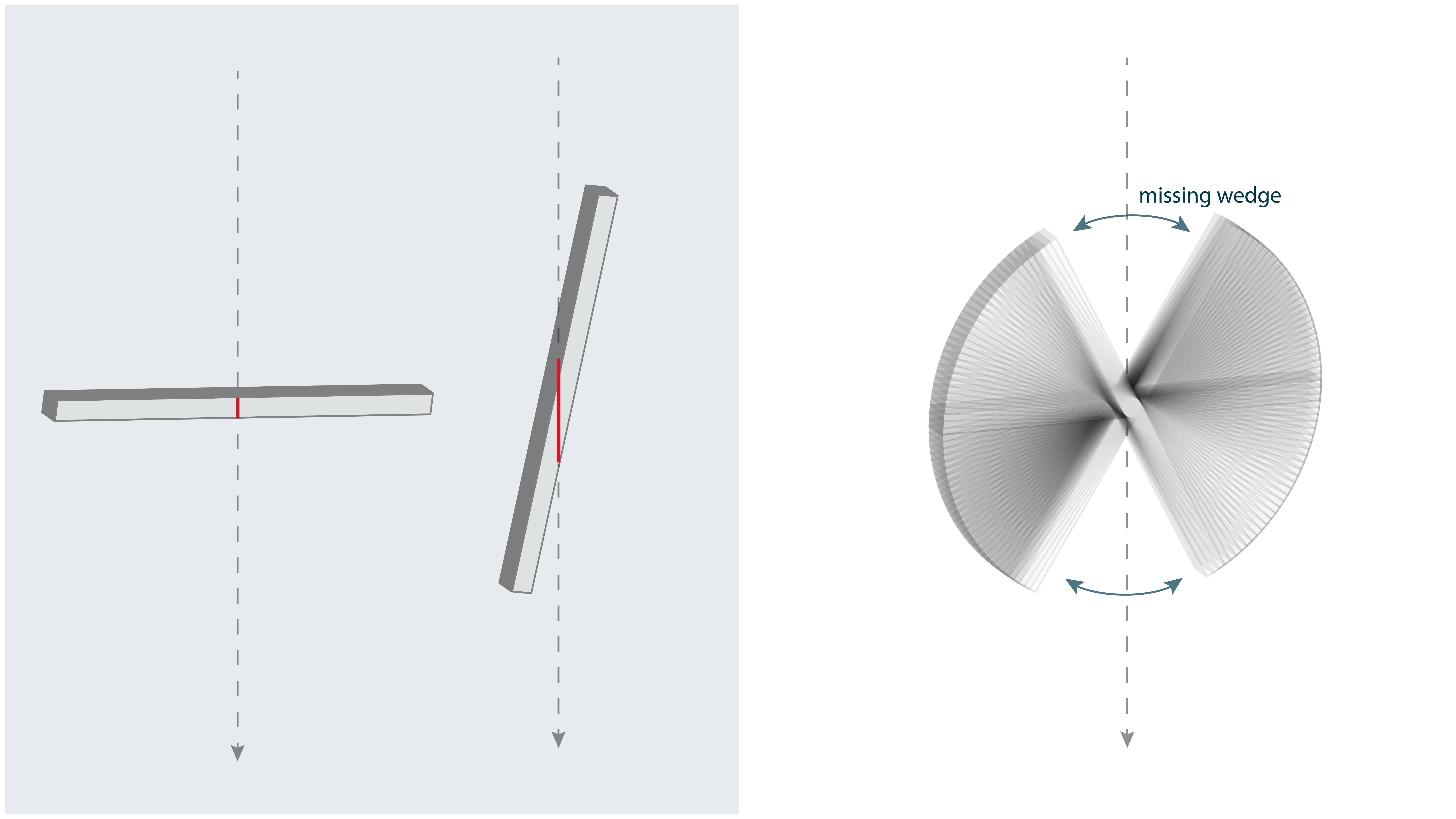
Think again about how a tilt-series is made: by taking images from different angles. If we could tilt the sample all the way to 90º, we would have information from every angle. But the sample gets effectively thicker as we tilt it, since the beam has to pass through more and more of the surrounding material. This effect, illustrated on the left, usually becomes prohibitive beyond 60º, so a typical tilt-series spans only ~2/3 of the possible angles, leaving a “missing wedge” of information corresponding to those high tilt angles, as you can see on the right. The missing wedge blurs densities in the direction of the imaging beam. In practice it means that if we look at a cross-section of a cell, we cannot trace thin features like membranes all the way around. You will see an example of this effect in Chapter 5.10.


