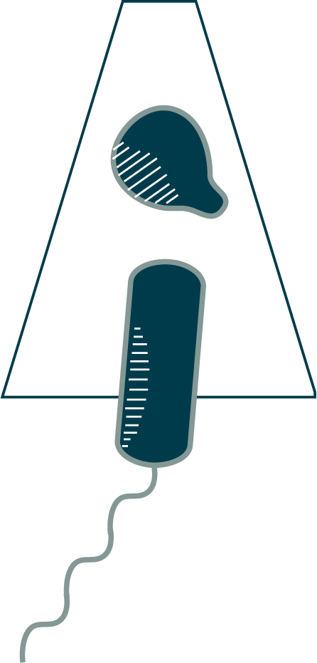
This C. necator cell is at a later stage of cytokinesis. In most cells, constriction would be more or less uniform around the circumference by now, but in some cells, including this one, the asymmetry persists.


Division can also be asymmetric in a different way: how constricted one side of the cell is relative to the other. You can see that in this Cupriavidus necator cell. Asymmetric constriction is most common early in division, but it sometimes persists longer (⇩). To understand why this happens, we need to take a closer look at how cells divide.
To divide, your cell’s membrane and cell wall constrict. But remember that the cell wall is resisting the non-trivial force of turgor pressure pushing outward. So how can it overcome this pressure and grow inward? For almost all bacteria and many archaea, the answer is a protein contractor called FtsZ (Filamenting Temperature-Sensitive mutant Z, named for a genetic screen for cells that failed to divide, and therefore grew into long filaments). FtsZ is another piece of your cell’s cytoskeleton, and is homologous to eukaryotic tubulin. FtsZ polymerizes into filaments that are linked to the cell membrane around the division plane (⇩). The filaments are highly dynamic, forming, disassembling and reassembling within seconds.
How does FtsZ find the right spot? There are multiple mechanisms, but an elegant one in cells like this involves an inhibitor that localizes to the cell poles, making a repressive gradient that is strongest at the ends of the cell and weakest in the middle. As the rod-shaped cell grows, the inhibitor concentration at the middle eventually drops low enough that FtsZ can polymerize. Most cells also inhibit FtsZ polymerization when the genome is still in the way, a mechanism called “nucleoid occlusion.” Some species do this with an FtsZ-inhibiting protein called MipZ that concentrates around the ParB chromosomal handle, ensuring that FtsZ filaments do not form until the chromosome is clear.
Once everything is ready (the cell is long enough and the nucleoid out of the way), FtsZ filaments form at the division site. Initially, a single, short filament (or a few) appears. Already, this is enough to begin constricting the cell, which explains why many bacterial cells, including this one, start to constrict on one side only. FtsZ filaments run parallel to the cell membrane, so they appear as dots in cross-section; if we rotate the cell around to look down its long axis, we can see them.
Note: we cannot see the membrane (and any associated FtsZ filaments) all the way around the cell due to the “missing wedge” of information in cryoET imaging. See Chapter 1.6 for an explanation.
This C. necator cell is at a later stage of cytokinesis. In most cells, constriction would be more or less uniform around the circumference by now, but in some cells, including this one, the asymmetry persists.
While the structure of FtsZ is known, as seen here from Staphylococcus aureus [45], its mechanism is not. It is an enzyme that hydrolyzes GTP, so one hypothesis is that the curvature of the filament changes as the chemical reaction drives subunits from one conformation to another. This different filament conformation may pull inward on the attached membrane, allowing the cell wall to build inward behind it.