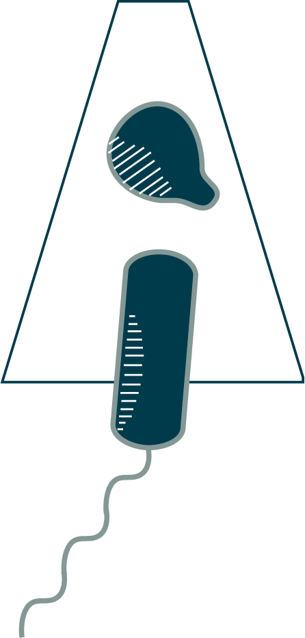
Why stop at one membrane? Eukaryotic cells use internal membranes to form specialized compartments like the nucleus and mitochondria. While bacteria lack such organelles, many create an additional compartment outside the cell with a second, outer membrane. Such bacteria, like the Hydrogenovibrio crunogenus cell you see here, are called diderms (“double skin”). The extra compartment between their membranes is known as the periplasm (“substance between”). This antechamber contains a unique subset of proteins, many of which function in escorting things into and out of the main cell.
Compared to the inner membrane, the outer membrane has some unique properties. It is more permeable and not proton-tight (so it cannot be used to generate ATP). It is also usually asymmetric, with very different molecules in the outer leaflet than in the inner (⇩). A few species of bacteria, particularly pathogens, have labile outer membranes (you will see an example in Chapter 8.2). Most, however, firmly anchor their outer membrane to the cell wall (⇩).
Rather than containing many layers of peptidoglycan like in the Listeria monocytogenes you just saw, the cell wall of diderms is usually composed of a single layer of peptidoglycan mesh (⇩), which is visible here as a thin line in the periplasm. This single-layered cell wall presents a considerable challenge for growth: how can you remodel it to grow bigger without letting turgor pressure burst the cell in the process? The answer, as we are beginning to figure out, is very carefully (⇩).
The difference in thickness of monoderm and diderm cell walls enables a well-known classification system: the Gram stain, which binds peptidoglycan. Gram-positive bacteria, typically monoderm, contain much more peptidoglycan than Gram-negative bacteria, which are typically diderm.